The fundamentals
Cancer and its generation is regulated at 4 crucial stages(locations).
Cell Cycle regulation
The retinoblastoma gene product example
cdk inhibitors
Signalling from outside the cell to the nucleus
Cytoplasmic response Elements
G-Protien linked receptors
Receptor tyrosine Kinases
Tyrosine Kinase Assosciated Receptors
Serine/threonine Kinase Receptors
Tyrosine Phosphatase Receptors
Cytoskeleton and cell adhesion
Regulation of cell death
Regulation of gene transcription
Cell Cycle regulation Go back
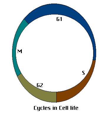
The cell undergoes a series of changes in its life, whether actively involved in proliferative activity, or quiescent. All the changes are tightly regulated and the transitions from one phase to the other are closely monitored.Phase M is the mitotic phase, new nucleii are formed and the cytoplasm is equally divided amongst the two daughter cells formed. Phase G1(gap 1) determines the readiness of the cell to commit to replication. Phase S is the phase of DNA synthesis. Here replication of the genetic material is carried out and further replication is held back. Phase G2 determines the fidelity of the replication of DNA and repairs are carried out.
Protien kinases whose activation depends on a cyclin protien, called cyclin dependant kinases regulate the transitions between the various phases. The enzymatic activity of the kinase is dependant on its assosciation with a cyclin and its phosphorylation state. Degradation of cyclins is carried out by the ubiquitin-proteasome complexes(during Phase M).
For example, the Cyclin B/cdc2 complex regulates the transition between G2 and M. An activated cdk in this system produces changes like
- Phosphorylation of Histone
- Phosphorylation of nuclear lamins
- Phosphorylation of microtubule assosciated protiens
leading to
- chromosome condensation
- breakdown of nuclear lamins
- spindle formation
respectively.
Cyclin B/cdc2 complex is frequently refered to as the Mitosis promoting factor(MPF).
The retinoblastoma gene product example
The retinoblastoma gene product designated Rb, is present normally in a hypophophorylated state. In this state, the molecule serves to scavenge E2F (a transcription factor) molecules. Free E2F can heterodimerize with other transcription factors like DP1 and directly signal the basal transcription machinery to start DNA replication. But when bound to Rb, DP1 is unable to activate the replication machinery(the activity of the E2F/DP1 is reduced by the Rb bound to DP1). Removal of Rb from DP1 by multiple phosphorylation at several pocket binding sites on the Rb releases free DP1 and transcription can proceed
In other words, Rb promotes differentiation while 'inhibition ' of Rb brings about transcriptional activity. Mulitple phosphorylation of the Rb molecule is carried out by two cdks-
- Cyclin D/cdk4 or cdk6
- Cyclin E/cdk2
Growth factors promote cdk activity, wheras TGFb, contact inhibition, cAMP inhibit cdk acrivity through various mechanisms.
Activity of the cdks in also modulated by cdki-cdk inhibitors
Usually designated as p(some number), eg; p21, inhibitors are low molecular weight protiens that inhibit cdks. Genetic alterations in cdki esp. p16 and p15 ahve been found very frequently in various tumors.
p53, a cdki is known as the guardian of the genome at the G1 checkpoint. It is expressed in case of extensive DNA damage. It can either induce p21 production to halt cell cycle progression or set of f an apoptotic pathway if the DNA damage is too great. Mutations in p53 are the most common genetic alteration found in human cancers. Mutations always seem to involve the region between codon 120 and 290. p53 is the target of many tumor causing viruses like
- human papilloma virus(protien E6 of the virus)
- Adenovirus E1B protien
- SV40 large T antigen
All seem to interfere with p53 function.
Codon 249 is mutated by the chemical substance aflatoxin, leading to hepatomas.
Signalling from outside the cell to the nucleus Go back
Signalling from inside the cell is carried out by a variety of receptor/intracellular domain moieties. These complexes, obvioulsy, have functions both in the resting phase of the cell and the division/differentiation arm of cell life progression. Different types or "approaches" are found in cells. These include
Cytoplasmic response elements
Example no 1.
The receptor for retinoic acid in cells is a intracytoplasmic element. Mutatiuon in the gene encoding for the receptor occurs in acute promyelocytic leukemia, the mutation being the result of the translocation t(15:17). It has been found that nuclear localization and proper activation of the retinoic acid receptor is needed for terminal differentiation of developing leukocytes(don't ask my how).
G-Protien linked receptors
G-Protien linked receptors are implicated in cancers in two basic pathways.
- Activation of CREBs through the Protien Kinase A pathways
- Inactivation of certain inhibitory molecules leading to translocation of intranuclear transcription factors through the Protein Kinase C pathway.
1. N.B I couldn't figure out the mode of action of cAMP response elements and CRE binding proteins. The pathway, as I saw it, was 'self-terminating'. If you are able to figure it out or know it already, do tell me.
- The more important pathway concerns the activation of protien kinase C.The basic framework of the regulatory pathway involving PKC starts with cAMP releasing intracellular calcium stores and bringing in extra cellular calcium. This leads to production of second messengers IP3 and DAG via the Phospholipase C pathway. DAG stimulates Protien Kinase C which activates a family of serine/threonine kinases which directly then act on factors regulating transcription. For eg.
DAG-mediated activation of Protien Kinase C
|
|
\/
Activation of the MAP Kinase pathway
|
|
\/
Phosphorylation of IkB(an inhibitor molecule, prevents an transcription acvivating heterodimer complex from reaching the nucleus)
|
|
\/
Release of the activating heterodimers(these belong to the rel family).
|
|
\/
Nuclear translocation of the activating heterdimers
|
|
\/
Transcription
Receptor tyrosine kinases
The receptors for growth factors like
- PDGF
- FGFs
- EGFs
- TGF(+)
- nerve growth factor
- heregulin
- insulin
- insulin-like growth factors
- Stem cell factors
- Colony stimulating factors
are all Receptor tyrosine kinase complexes.
A brief note on the structure of these receptors
Receptor tyrsoine kinase complexes are simple in structure as compared to the G-protien receptor complexes. But the activation of intracellular events by these receptors take as sinuous a path as is the case of the G-Protien transducer elements.
[image not archived: images/Tyr_kin.gif]
Ligand binding induces dimerization or oligomerization of the receptor subunits leading to activation of the tyrosine kinase activity on the intracellular domains and susequent autophosphorylation of certain segments on the intracellular chain. These phosphorylated segments then act as binding sites for other signal transduction molecules. Many enzymes (like the nonreceptor tryosine kinase src) bind to these phosphorylated binding sites and get activated. Some protiens serve as docking proteins exclusively in that , they lack enzymatic activity but they bind to the binding sites and subsequently activate other enzymes, serving as bridges. The docking protiens bind to the binding sites on the receptor using SH2, SH3 or PH domains and activate downstream events.
Tyrosine Kinase receptors have been shown to be involved in the activation of the
ras
fos/jun/AP-1 family of transcription factors
c-myc activation
Overexpression of tyrosine kinase receptors has been seen in many cancers,
-lung
-bladder
-head and neck
breast
ovarian cancers.
Tyrosine kinase assosciated protiens
The receptors for
-growth hormone
-prolactin
erythropoeitin
-IL-2, IL-3, IL-4, IL-6, IL-7
-granulosyte CSF
interferon gamma
are all tryosine kinase assosciated receptors.
The kinases assosciate with the receptors by docking proteins. to mainly, three classes of kinases
the src family of kinases
The Janus family of kinases
The syk family of kinases
and lead to similar types of signalling cascades as mentioned above. For eg, the Janus family of kinases activate a class of molecules called STATs(signal transduction and activation).
These receptors are frequently overexpressed in tumours of hematopoietic origin.
Serine/Threonine Receptor Kinases
These receptors recognise TGFb and bone morphogenetic factors. TGFb promotes cell differentiation by induction of cdki. Loss of expression of TGFb receptors in seen in colon carcinoma and lymphomas
Tyrosine Phospatase Receptors
CD45 on T-cells is the prototype here. T-cell activation involves removal of a phosphate residue on lck , a kinase of the src kinase family. This activation of lck leads to signal transduction through the T-cell antigen receptor. It seems that activation of lck depends on the activation of the CD45 receptor on the T-Cell surface. Role of CD45 in neoplasia in not known.
Cytoskeleton and cell adhesion Go back
Three major types of fibres make up the cytoskeleton of normal cells: Actin, microtubules and intermediate filaments. Microtubules are polymers of tubulin, form transport promoting railways inside the cell. Actin contrbutes to cell motility and is a crucial component of cell-to-cell junctions and focal contacts(cell-to-matrix). There are many types of intermediate filaments like keratin, vimentin, desmin etc. Thy give the cell tensile strength and form components of desmosomes and hemidesmosomes.
In addition normal cells maintain cell-cell contact through calcium-dependant protiens called cadherins, cells involved in host defense use homologous protiens called selectins.
Cellular cytoskeletal disorganization is a hallmark of cancer. (It looks as though the cell is getting ready to metastazise--Ed.)
The adenomatous Polyposis Coli Gene Product example
This is an example where a cytoskeletal regulatory element has been implicated the pathogenesis of a cancer. The Apc gene product is defective in familial polyposis. The wnt protien, a matrix bound ligand activates the APC gene product. Activated APC (made active by phospjhorylation) sequesters beta-catenin and reduces its activity in the cell. This function of APC is impaired in familial polyposis and beta-catenin is set free, leading to disruption of adherens junctions and other cytosekeletal changes.
Regulation of cell death Go back
"Maybe you should cut some root to make the tree survive.
Some cells must die to preserve the rest of the organism "
Apoptosis is a form of cell death wherein intre- of extracellular events trigger the activity of a family of enzymes which degrade nuclear DNA. For example, cell signals through the TNF receptor leads to the activation of enzymes in the ICE (IL-1 conveting enxyme) family and ICEs cleave certain protiens at specific asparagine residues and initiate the cell death pathway. This pathway has been implicated in the killing of autoreactive lymphocytes.
The bcl-2 family of genes modulate apoptosis. Members like bcl-2, bcl-Xl prevent cell death, members like bax, bad, bak, promote cell death. bcl-2 family members can homo- or heterdimerize among themselves , the combinatorial effects allow a fine degree of control over cell survival.
When any of the death promoters exist as homo- or heterodimers, the cell dies. When death promoter dimerizes with death preventers like bcl-2 death is provented. Thus relative levels of promoters and preventers determine cell life.
Abnormal expression of bcl-2 (due to t(14:18), the bcl-2 being present on 18q) is unequivocally linked to follicular lymphoma.
Abnormal levels of bcl-2 in the cell can prevent the p53 mediated cellular death pathways and thus allow proliferation of cells with extensive DNA damage, thereby forming the basis of cancer pathogenesis. bcl-2 seems to confer drug resistance and radioactive therapy refractoriness in a similar fashion.
Regulation of gene transcription
Please refer Harrison's Vol-1 for text(sorry guys).
Cancer Genetics-The Fundamentals Go back